
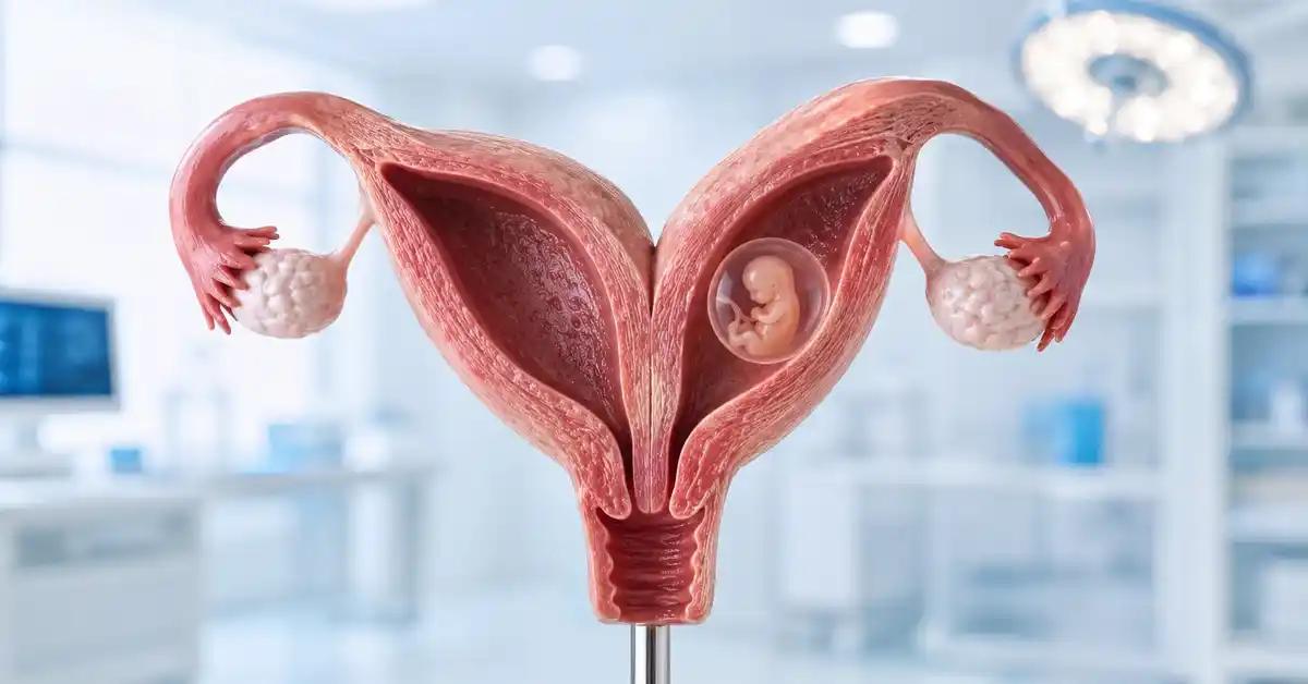
Congenital Uterine Anomalies And Their Effect On Pregnancy

Manar Hegazy

Majd Eddin Khaled
Congenital uterine anomalies are differences in the shape or development of the uterus that form before birth. Many women do not know they have one until they experience delayed pregnancy, recurrent miscarriage, preterm birth, or a detailed pelvic ultrasound. Some anomalies are mild and have little impact, while others may increase the risk of pregnancy loss or obstetric complications.
A uterine anomaly does not automatically mean that pregnancy is impossible. Many women with a differently shaped uterus conceive and deliver successfully. However, when there is recurrent miscarriage, implantation failure, preterm birth, or infertility, evaluating the uterus becomes important. At Fertiliv, the diagnosis is interpreted carefully: What type of anomaly is present? Does it affect the uterine cavity? Is treatment needed before pregnancy or embryo transfer?
What Are Congenital Uterine Anomalies?
Congenital uterine anomalies develop when the Müllerian ducts do not form, fuse, or resorb normally during fetal development. These ducts form the uterus, fallopian tubes, and the upper part of the vagina. A difference in this process can result in a uterus with an unusual shape or internal cavity.
The anomalies range from mild variations, such as an arcuate uterus, to more significant forms such as septate uterus, bicornuate uterus, didelphys uterus, or unicornuate uterus. Their impact varies, so the exact diagnosis matters more than simply saying that the uterus is “abnormal.”
Are They Common?
They are not extremely rare, but many remain undiagnosed because they do not always cause symptoms. They may be found during fertility testing, after miscarriages, after preterm birth, or during imaging such as HSG or 3D ultrasound.
Not every anomaly requires treatment. Some need only observation. The decision depends on the type of anomaly and the patient’s pregnancy history.
Do They Cause Infertility?
Some uterine anomalies may not prevent conception but may affect implantation or pregnancy continuation. A woman may conceive but experience miscarriage, preterm delivery, or abnormal fetal position. In other cases, the anomaly is not the main cause of infertility.
This is why the full fertility picture should be evaluated: ovulation, ovarian reserve, fallopian tubes, semen analysis, endometrium, and hormones. A uterine anomaly should not automatically be blamed for every fertility issue.
Types Of Congenital Uterine Anomalies
Several main types exist. A septate uterus contains a partition inside the uterine cavity. A bicornuate uterus has a deep external division at the top of the uterus. A didelphys uterus has two uterine cavities and often two cervices. A unicornuate uterus is smaller and develops mainly from one side.
The reproductive impact differs by type. A septate uterus is often discussed in recurrent miscarriage. A bicornuate, didelphys, or unicornuate uterus may be more associated with preterm birth, abnormal fetal position, or pregnancy monitoring concerns.
Septate Uterus
A septate uterus occurs when an internal septum remains inside the uterine cavity. This septum may be less suitable for implantation or early pregnancy support. It is one of the anomalies most often linked with recurrent miscarriage.
If recurrent miscarriage or repeated implantation failure occurs with a clear septum, hysteroscopic septum resection may be discussed. However, treatment should be based on accurate diagnosis and the patient’s history, not on imaging alone.
Bicornuate And Didelphys Uterus
A bicornuate uterus develops when the upper part of the uterus does not fuse completely, creating two horn-like areas. Pregnancy can occur, but there may be increased risk of later pregnancy loss, preterm birth, or breech presentation.
A didelphys uterus means two separate uterine cavities, often with two cervices and sometimes a vaginal septum. Many women can carry pregnancies, but monitoring may need to be more careful. Surgery is not automatic and is reserved for selected cases.
Read about: Weak Ovulation: A Clear Guide to Diagnosis and Treatment Options
How Uterine Anomalies Affect Pregnancy
The effect depends on how much the uterine cavity is altered, the available space for the pregnancy, blood supply at the implantation site, and endometrial quality. If the cavity is divided or distorted, implantation may occur in a less favorable area, increasing the chance of loss.
Some anomalies may also affect fetal position in later pregnancy. Breech or transverse presentation may be more common in certain uterine shapes, and the risk of preterm delivery may be higher.
Recurrent Miscarriage
Septate uterus is the classic anomaly considered in recurrent miscarriage. If an embryo implants on the septum, the blood supply may be less supportive, and pregnancy may stop developing. This makes uterine cavity assessment important after repeated losses.
However, recurrent miscarriage is not always uterine. Chromosomes, hormones, antiphospholipid syndrome when indicated, egg quality, and sperm quality should also be reviewed.
Preterm Birth And Fetal Position
Bicornuate, unicornuate, and didelphys uteri may be associated with a higher risk of preterm birth or abnormal fetal position. This may be related to uterine shape, size, or how the uterus expands during pregnancy.
If pregnancy occurs, monitoring may include cervical length assessment, fetal growth follow-up, and attention to fetal presentation later in pregnancy.
Read about: Endometriosis And Pregnancy: Is Natural Conception Possible Or Is IVF Necessary?
Uterine Anomalies And Infertility
Some women discover a uterine anomaly during infertility evaluation. The important question is whether the anomaly is truly affecting fertility. If the uterine cavity is distorted, implantation may be affected. If the anomaly does not significantly affect the cavity, another cause may be more likely.
Fertility care should not focus only on the uterus. Semen analysis, ovulation, ovarian reserve, tubes, and general reproductive health must also be reviewed.
Implantation Failure
When embryo transfers fail repeatedly, especially with good-quality embryos, the uterine cavity should be assessed carefully. A septum, adhesions, polyps, or subtle cavity issue may not be visible on basic ultrasound.
3D ultrasound or hysteroscopy may help when there is a clear indication. However, surgery should not be performed without a specific reason.
IVF/ICSI And Uterine Anomalies
IVF/ICSI can help with male factor infertility, blocked tubes, low ovarian reserve, or other causes, but it does not change the shape of the uterus. If the uterine cavity is affected, this should be considered before embryo transfer.
In some cases, embryos may be created and frozen first, while the uterus is evaluated or treated before transfer. This protects embryo opportunities while allowing time for safer planning.

How Are Uterine Anomalies Diagnosed?
Diagnosis requires accurate imaging because some anomalies can look similar at first. Standard 2D ultrasound may provide an initial impression, but 3D ultrasound is often more useful for assessing both the uterine cavity and the outer shape of the uterus. In selected cases, MRI or hysteroscopy may be needed.
Distinguishing septate uterus from bicornuate uterus is especially important because treatment differs. A septum is inside the cavity and may be treated hysteroscopically in selected cases. A bicornuate uterus involves the external uterine shape and is not treated the same way.
3D Ultrasound
3D transvaginal ultrasound is one of the most useful tools for assessing uterine anomalies. It can show the cavity and outer uterine contour more clearly than standard ultrasound. This helps determine whether the issue is an internal septum or an external division.
It is relatively non-invasive and may reduce the need for more invasive tests in many cases. Interpretation should be done by an experienced clinician.
Hysteroscopy And MRI
Hysteroscopy allows direct visualization of the uterine cavity and can be used for treatment if a septum, adhesion, or polyp is present. However, hysteroscopy alone does not fully show the external uterine contour, so 3D ultrasound or MRI may be needed.
MRI is useful in complex cases, unclear imaging, or when cervical, vaginal, or renal-associated anomalies are suspected. The choice of test depends on the clinical question.
Read about: Uterine Fibroids And Fertility: When Do They Affect Pregnancy And How Are They Treated?
Do All Uterine Anomalies Need Treatment?
No. Not all congenital uterine anomalies require treatment. Some are found incidentally and do not cause significant problems, especially if the woman has had a normal pregnancy. Observation may be enough.
Treatment is considered when the anomaly clearly affects the uterine cavity or is associated with recurrent miscarriage, repeated implantation failure, or pregnancy complications. The potential benefit must be balanced against the risks of intervention.
Treatment Of Septate Uterus
Hysteroscopic septum resection may be discussed in women with recurrent miscarriage or infertility when a significant septum is confirmed. The procedure is usually performed through the cervix without abdominal surgery and aims to create a more regular uterine cavity.
Accurate diagnosis is essential. Mistaking a bicornuate uterus for a septate uterus can lead to inappropriate treatment.
When Observation Is Enough
Observation may be enough when the anomaly is mild, does not significantly distort the cavity, and there is no history of miscarriage, preterm birth, or repeated implantation failure. Some bicornuate or didelphys uteri may also be managed conservatively if pregnancy outcomes are acceptable.
Not every abnormal image needs surgery. Good medicine means treating only when treatment is likely to help.
Read about: Chronic Diseases And Fertility: How Can You Achieve Parenthood Despite Health Challenges?
Associated Conditions To Consider
Because the reproductive tract and urinary system develop closely during fetal life, some uterine anomalies may be associated with kidney or urinary tract differences. This is especially relevant in unicornuate uterus or more complex anomalies.
Some anomalies may also be associated with a vaginal septum or cervical differences. These can affect menstruation, intercourse, procedures, or delivery planning. A complete gynecologic evaluation is therefore important.
Kidney Evaluation
Not every patient needs kidney imaging, but it may be reasonable in selected anomalies. A renal ultrasound may be requested when a unicornuate uterus, didelphys uterus, or complex anomaly is diagnosed.
This is not only about fertility. It helps protect the patient’s general health and plan pregnancy or surgery safely.
Vaginal Septum
A vaginal septum may occur with didelphys uterus or complex Müllerian anomalies. It may cause pain, difficulty with intercourse, menstrual obstruction, or delivery concerns. Treatment depends on symptoms and reproductive plans.
If it causes no symptoms, observation may be enough. If it affects quality of life or pregnancy care, treatment may be discussed.
Read about: Blocked Fallopian Tubes: Is It The End Of The Motherhood Dream?
Pregnancy With A Congenital Uterine Anomaly
Pregnancy can succeed with a uterine anomaly. The level of monitoring depends on the type of anomaly and previous history. The doctor may follow cervical length, fetal growth, fetal presentation, and signs of preterm labor more closely.
Cesarean delivery may be more common in some cases because of fetal position or uterine shape, but it is not automatic for every patient. Delivery planning depends on the actual pregnancy course.
Cervical Monitoring
Some patients with uterine anomalies may need cervical length monitoring, especially if they have a history of preterm birth or later pregnancy loss. If cervical shortening is found, the doctor may discuss progesterone or other management depending on the case.
Early monitoring helps identify risks before symptoms appear.
Fetal Position And Delivery
Because uterine shape may affect fetal movement and space, breech or transverse position can be more common in some anomalies. If the baby is not head-down near delivery, cesarean birth may be more likely.
The final delivery plan depends on fetal position, maternal health, fetal growth, placenta, and cervical conditions.
Read about: Premature Ovarian Insufficiency (POF): Causes, Symptoms, And Medically Available Fertility Options
Fertiliv’s Role In Uterine Anomaly Evaluation
Fertiliv evaluates congenital uterine anomalies as part of fertility and pregnancy planning. The assessment begins with history: infertility, miscarriages, preterm birth, failed embryo transfers, unusual period pain, or previous imaging findings. Then the appropriate test is selected, such as 3D ultrasound, hysteroscopy, or MRI when needed.
After diagnosis, the type and likely impact are explained. If treatment may help, it is discussed clearly. If treatment is not needed, unnecessary intervention is avoided.
Before IVF/ICSI
Before embryo transfer, the uterine cavity should be suitable. If a septum or cavity problem is present, treatment before transfer may be more logical than placing a good embryo in an unsuitable environment. In some cases, embryos can be frozen while the uterus is prepared.
This planning protects embryo chances and reduces avoidable risk.
A Plan Based On The Type
A septate uterus is not the same as a bicornuate uterus. A unicornuate uterus is not the same as an arcuate uterus. The plan must depend on the type, severity, age, pregnancy history, and fertility goals.
Fertiliv uses individualized planning to avoid both over-treatment and under-treatment.
Read about: Enlarged Uterus: Its Effect on Fertility and the Latest Treatment Options
Conclusion
Congenital uterine anomalies can affect pregnancy in different ways. Some are mild and need no treatment, while others may be linked with recurrent miscarriage, implantation failure, preterm birth, or abnormal fetal position. Accurate diagnosis is essential, especially to distinguish septate uterus from bicornuate uterus because treatment differs.
Not every anomaly requires intervention. Treatment is considered when the uterine cavity is significantly affected or when the patient has miscarriage, implantation failure, or pregnancy complications that may be related to the anomaly. Fertiliv helps patients understand the diagnosis, evaluate the real impact, and choose the safest plan before pregnancy or embryo transfer.
Frequently Asked Questions: Congenital Uterine Anomalies And Their Effect On Pregnancy
Does A Uterine Anomaly Always Prevent Pregnancy?
No. Many women conceive and deliver with uterine anomalies, but some types may increase miscarriage or preterm birth risk.
Which Uterine Anomaly Is Most Linked To Miscarriage?
Septate uterus is one of the anomalies most often discussed in recurrent miscarriage.
How Are Uterine Anomalies Diagnosed?
They are often diagnosed with 3D ultrasound, and sometimes hysteroscopy or MRI is needed.
Should Every Uterine Septum Be Removed?
No. The decision depends on septum size, cavity effect, miscarriage history, and implantation history.
Can IVF/ICSI Be Done With A Uterine Anomaly?
Yes, but the uterine cavity should be assessed before embryo transfer. Treatment or delayed transfer may be needed in selected cases.